Rating of the best fluorographs for 2025

Fluorography is considered one of the most popular types of research. An X-ray image will help to identify many diseases and choose the right course of therapy. This method will be able to diagnose tuberculosis, pneumonia, neoplasms and other pathologies. With the help of fluorographic equipment, not only the lungs, but also other organs of the human body are examined.
From this review, buyers can learn about the device, the principle of operation of fluorographic equipment, and which model is better to purchase. The rating takes into account user reviews. Based on them, the best brands and brands are selected.
Content [Hide]
Functions and types
The unit is necessary for making X-ray images of human organs. They show pathological changes.
This method was discovered by scientists from Italy and America. There are two types of studies: small and large frames.
The main organs for study are the chest, skeletal system and mammary glands. Devices are divided into the following types:
- stationary, made for working rooms of clinics and hospitals;
- mobile, used as mobile stations.
Types of devices:
- film devices. These devices are used less and less in medical institutions, as they are obsolete. Their work requires a special film and a laboratory. During the examination of the human body, X-rays pass through a specific organ.
- Digital fixtures. The principle of operation is in many respects similar to the film analogue. However, the rays that have passed through the body are reflected differently. The information is fed to a re-radiating screen, which converts the picture into a digital format. Modern units have many functions, so pictures are taken very quickly. Devices process information more fully. High-quality material is easier to analyze. A big plus is that the picture can be quickly sent by e-mail. In some cases, this is necessary for a quick analysis. In addition, the picture after the transfer does not lose its quality. Another significant advantage can be attributed to the minimum dose of radiation.
Film-type devices are gradually becoming obsolete. They were replaced by digital X-ray equipment. It has the following advantages:
- images are much easier to work with;
- pictures are displayed on the screen, they can be printed, sent on the network and stored in the base memory;
- irradiation is much less than from a film aggregate;
- minimal cost of consumables.
The digital diagnostic method is divided into 2 types. The first involves the use of a fluorescent monitor. In this case, a CCD matrix is used instead of X-ray film. In the second technique, a fan-shaped beam is used, which gradually scans the human organ under study. This method produces a small amount of radiation. However, it will take more time to get the final result.
Basic operating principles
All fluorographic units are equipped with special detectors that detect x-rays. The device together with the emitter and the collimator moves in the vertical direction. It distributes X-ray beams of light. The collimator produces fan-shaped radiation. During the passage of the beam through the human body, it penetrates into the window of the detector chamber.
Information from the registration system is transferred to the computer memory block. The digital image is formed in the form of a 1024x2048 matrix. The data is collected after the X-rays pass through the human body. The picture image appears on the screen about 10 seconds after the scanning process is completed.
Recommendations for choosing a device
Currently, manufacturers offer a wide range of equipment for X-ray rooms.
Parameters to pay attention to when choosing equipment:
- Type of feeding device. Such devices are of medium and high frequency range. They differ in the ripple of the anode voltage. All this prolongs the life of the X-ray tube and significantly reduces the amount of radiation for the body.
- Power supply unit. The higher this indicator, the better the image quality and the lower the beam component.
- Optimal contrast. A small indicator indicates a small dose rate.
- The aggregate must have a high URI input field. This option will make research more qualitative.
- The dynamic range must have the necessary breadth. This will ensure the transfer of halftones in fabrics of different types.
- Photo exposure meter. This device is necessary for taking pictures in automatic mode.
- Focal spots should be as small as possible. This affects the clarity of the image.
Selection criteria and price
You can buy equipment in special firms or order via the Internet. In stores that operate online, you can consult. Consultants will help you choose the best option that meets all the requirements of the user.
The price of new devices starts from 3,000,000 rubles.
You can purchase a universal fixture. In this case, you need to look at mobile complexes. They are designed both for work in the field and in stationary mode. Many medical institutions acquire models designed for different positions of the human body (sitting, lying, standing). This is necessary to provide diagnostics for patients with limited mobility. It is very important that the equipment has sufficient speed and is suitable for repair.
Rating of fluorographs for 2025
The list includes units that diagnose not only human chest pathologies, but also models that have the functions of testing the state of other organs. The rating includes mammography fluorographs and devices for recognizing pathologies of the skeletal system.
mobile
Where there are no diagnostic centers, mobile units are simply irreplaceable. Mobile chest stations are of great importance.
Italray Clinomat
votes 7
votes 7
It costs from 6,000,000 rubles.
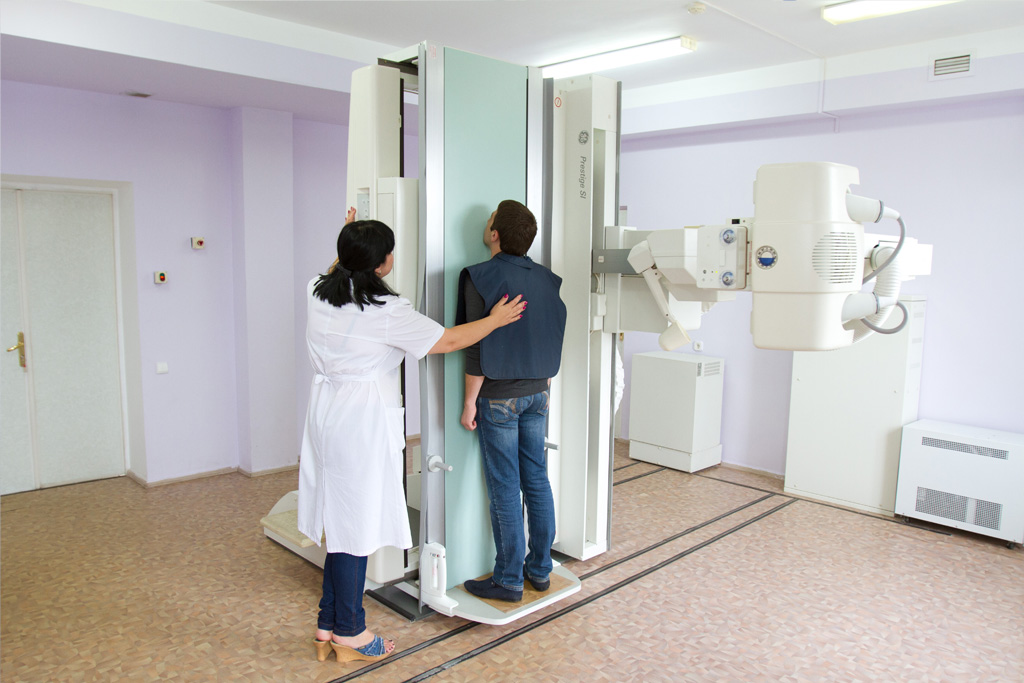
This is an open device. It diagnoses the human body in an upright position. The device has a high speed, correct laying and high repeatability. Diagnosis detects tuberculosis at the earliest stages. This is facilitated by high image quality.
The equipment allows you to make digital pictures of different organs. The unit can move. If it is installed on a car, then it will be possible to conduct surveys of people in the field.
The equipment is popular and is in the ranking due to the following devices:
- high-frequency inverter installation, which is controlled by the latest microprocessor;
- a high-speed tube equipped with a moving composite anode;
- vertical position of the patient;
- a tripod that performs the functions of placing an x-ray tube relative to the human body.
The unit can come in different configurations. The presence of options affects the characteristics of the device. Before buying, be sure to consult with the manufacturer's specialists.
Advantages:
- the device can be used as a mobile station;
- the patient is in an upright position;
- the unit has high performance;
- you can examine not only the lungs, but also other organs;
- The device is manufactured using innovative technologies.
Flaws:
- rather high price;
- there is no open access to lists of characteristics.
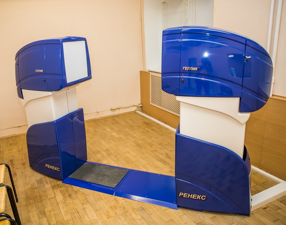
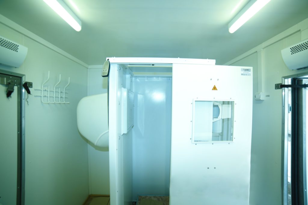
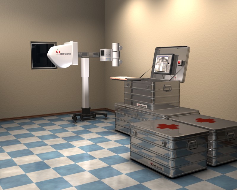
FMC S.P. Gelpik (in laying)
votes 9
votes 9
Average price: 5.5 million rubles.
These low-dose fluorographs fit easily into a box. They are used in various medical institutions. The main purpose of the fluorograph is to examine a large volume of patients in the field.With the help of the device, diseases such as tuberculosis, tumors, benign and malignant, etc. are detected. During the examination, a diagnosis is made. The device is easy to transport thanks to the box, where each part has its own place. The packaging prevents the fluorograph from being damaged during transportation, as well as getting wet in the rain.
Digital MF has its own features of work:
- thanks to the latest technologies, during the examination, patients receive the slightest radiation exposure, but the results are highly accurate;
- the patient during the examination is in a vertical position, the picture is taken in two projections - lateral and direct;
- the resulting digital images are processed and archived, and you can also get a hard copy of the picture;
- allows you to maintain a database of medical data in digital form;
- works in a single-phase network with a voltage of 220 volts, fluctuation in the region of 10% is permissible, the frequency should be 50 Hz;
- when it is not possible to connect to an external source, an autonomous station is suitable for operation, giving a voltage of over 4.5 kW.
Stationary devices are intended for examinations of patients in polyclinics and hospitals. They allow for the prevention of diseases and timely detection of the presence of pathological processes.
Advantages:
- can be transported;
- registration certificate available;
- clear pictures;
- convenient to store and move;
- can detect a wide range of diseases;
- connects to remote access on the network;
- has a large memory for examination results.
Flaws:
- the weight;
- personnel are exposed to radiation exposure;
- high cost.
Stationary
FC-Proton
votes 5
votes 5
The average cost is 5 million rubles.
The device promptly transmits data on the results of the examination.Previously created screenings remain in the memory of the device, with various diagnostic information. It is noteworthy that a radiologist and an X-ray laboratory assistant can work on a local computer network, each at their own place. Designed to examine a large number of people. It can be used to diagnose tuberculosis, cancer and other pathologies of the pulmonary system. It can be used in clinics for various purposes for reasons:
- patients receive ultra-low doses of radiation;
- you can change the focal length;
- the presence of an additional examination function with a resolution exceeding 4.2 lp / mm;
- excellent throughput allows you to take 60 shots per hour;
- full automation of processes providing calibration and self-diagnostics.
Advantages:
- you can do examinations of various organs and systems: lungs, ribs, thoracic spine and main blood vessels;
- allows you to connect via remote access;
- introduction of advanced technologies.
Flaws:
- the device is difficult to transport;
- radiation load;
- high price tag.
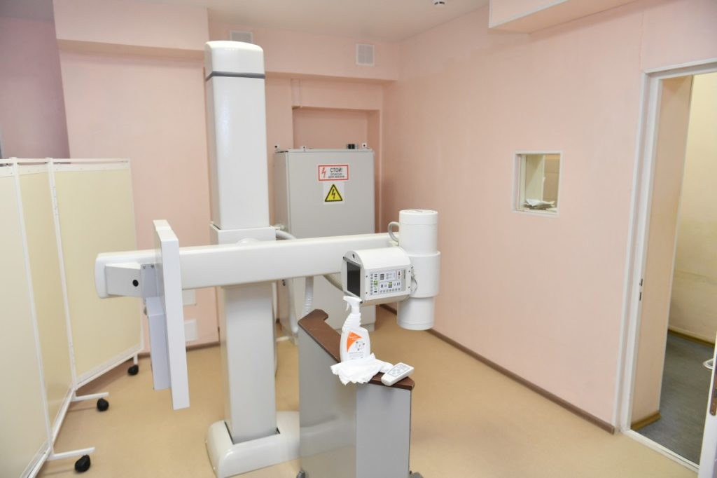
ProGraph-4000
votes 7
votes 7
The average cost is 4 million rubles.
ProGraph-4000 can be used to examine patients in standing and sitting positions. The device's camera is based on a CCD matrix. The device is equipped with DICOM 3.0 software, the results are sent to the printer of the same brand. Patients during the examination can either stand, sit or lie down. The device operates from a three-phase electrical network with a voltage of 380 V ± 10% with a resistance of up to 1 Ohm. The quality of the device is confirmed by a certificate. The device is in high demand due to:
- spatial resolution of 2.5 pl/mm, due to the geometric increase, the indicator reaches 3 pl/mm;
- rotating camera, thanks to which you can take pictures from different angles;
- a table transparent for the X-ray machine, which makes it possible to examine bedridden patients;
- that corresponds to the internationally recognized standard "DICOM 3.0";
- the possibility of automatic settings for various organs.
Advantages:
- the device takes pictures in different positions of the patient;
- an automated system helps the doctor to do examinations as quickly as possible;
- 1 shot per minute;
- advanced working methods;
- software that works according to world standards.
Flaws:
- dimensions;
- radiation load on employees;
- not transported.
 <
<
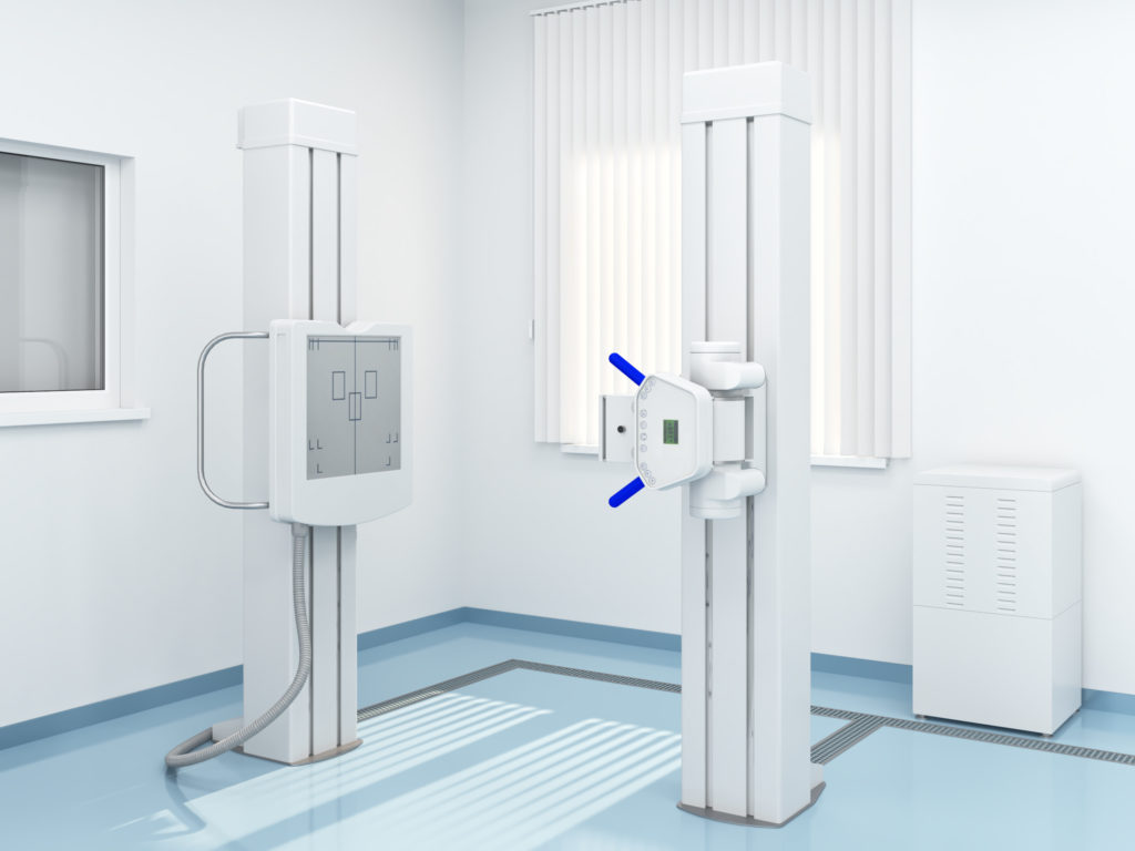
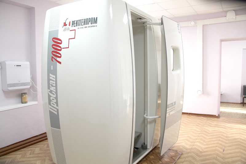
ProScan-7000
votes 9
votes 9
The cost is 5.2 million rubles.
This device is manufactured by the company "Rentgenprom". Used for examination in a standing position. The purpose of his work is the diagnosis of chest problems. 2 types of projection - front and side. With the help of it, verification studies are done in outpatient and inpatient departments.
The cabin is designed in such a way that the medical workers servicing the device receive the minimum dose of radiation. The unit can be connected to the mains via an adapter. The kit also includes a monitor, which the radiologist can use to monitor the correct position of the patient. In the kit with the device itself are:
- X-ray protective booth;
- scanning device;
- a linear silicon detector that is so durable that it does not need maintenance;
- high-frequency battery that can be connected to the network.
A workplace has been prepared for a specialist working with ProScan-7000, which includes a system unit, a drive for reading and writing disks, a monochrome monitor, a special SONY-UP 990 AD printer, a laser printer, and software for controlling the device.
Advantages:
- doctors do not receive radiation;
- a ready-made workplace for a laboratory assistant and a doctor;
- Hi-tech;
- works quickly;
- has all quality certificates.
Flaws:
- dimensions;
- examination only while standing;
- does not transform for movement;
- high price.
With the help of early diagnosis, you can quickly start treatment. To increase the chances of recovery, it is important to identify the pathology at an early stage. Therefore, you need a fluorograph that will take clear pictures and work quickly. The rating included the best representatives made on the basis of advanced technologies.
new entries
Categories

The best men's sneakers in 2025
Views: 124034

The Best Complex Vitamins in 2025
Views: 121940

Rating of the best steam inhalers for 2025
Views: 1199

Ranking of the best sugar substitutes for 2025
Views: 6091

The best playgrounds in Moscow in 2025
Views: 13695

Rating of the best waterproofing for 2025
Views: 3430
Useful

Ranking of the best vacuum sealers for 2025
Views: 1937

Rating of the best electric hobs for 2025
Views: 2341


Best Antivirals in 2025
Views: 33334

Top 15 hoverboards of 2025. Decent value for money
Views: 29339
Popular Articles
-

Top ranking of the best and cheapest scooters up to 50cc in 2025
Views: 131652 -

Rating of the best soundproofing materials for an apartment in 2025
Views: 127691 -

Rating of cheap analogues of expensive medicines for flu and colds for 2025
Views: 124519 -
The best men's sneakers in 2025
Views: 124034 -
The Best Complex Vitamins in 2025
Views: 121940 -

Top ranking of the best smartwatches 2025 - price-quality ratio
Views: 114980 -

The best paint for gray hair - top rating 2025
Views: 113396 -

Ranking of the best wood paints for interior work in 2025
Views: 110319 -

Rating of the best spinning reels in 2025
Views: 105330 -

Ranking of the best sex dolls for men for 2025
Views: 104367 -
Ranking of the best action cameras from China in 2025
Views: 102217 -

The most effective calcium preparations for adults and children in 2025
Views: 102012