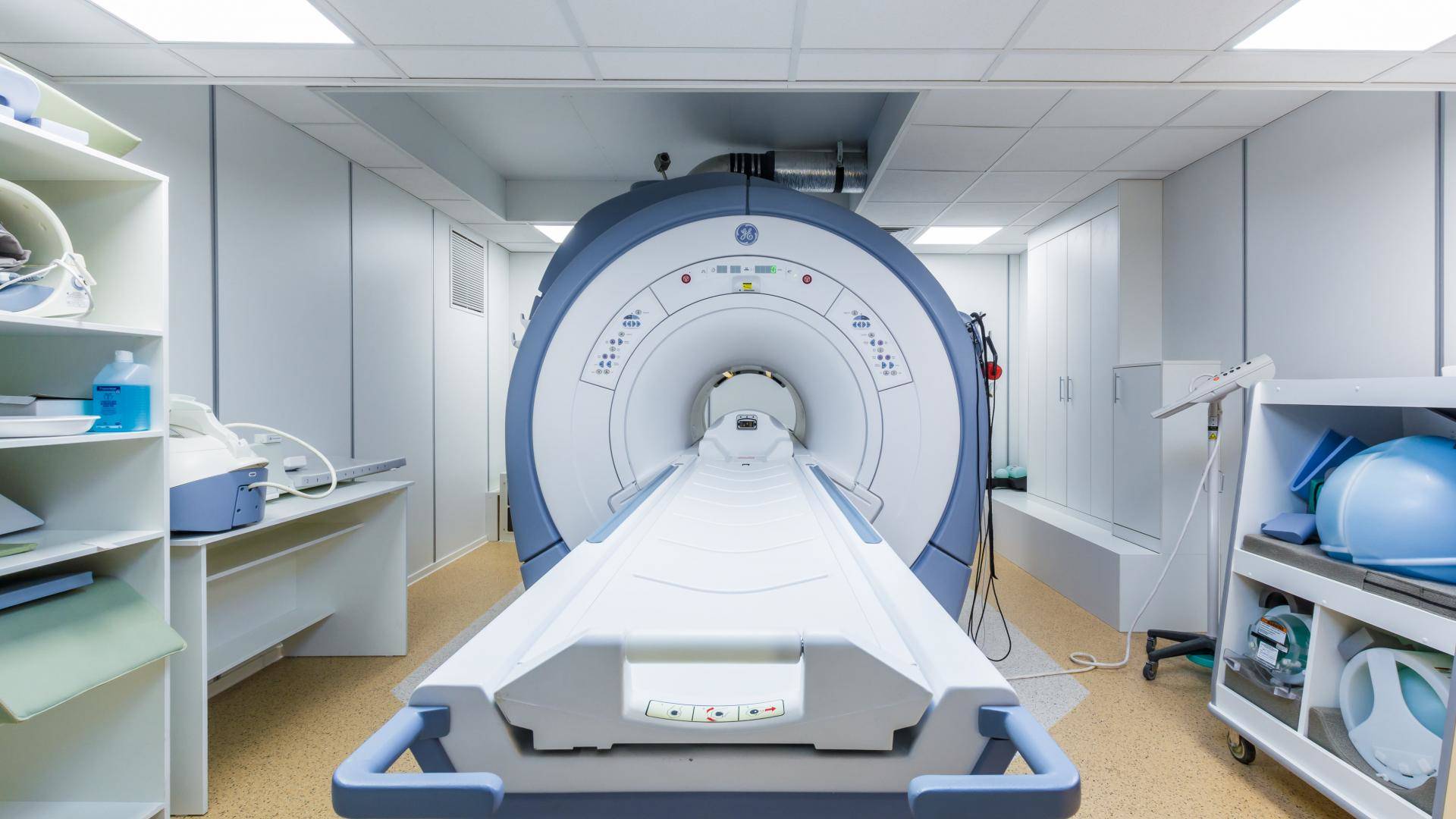
Rating of the best MRI machines for 2025
The development of computer technology and medical equipment has recently improved the quality of patient examination. Of the large number of devices for diagnosing the state of human health, one of the leading places is occupied by a magnetic resonance imaging (MRI), which allows you to assess even minor damage to bone and muscle tissue. The principle of operation of the device is to apply high-power magnetic radiation to the human body, which activates the movement of hydrogen particles, the location and speed of movement of which can determine the state of tissues, bones, and blood vessels.
As a result of numerous studies, the safety of this type of research has been proven. Many patients are afraid to undergo such a diagnosis, believing that it can cause the growth of neoplasms, but this is not true. Doctors note that diagnostics using a tomograph makes it possible to detect even defects in the initial stage, since the image is taken with high detail.
In this article, we will tell you how to choose such a device, what to look for in order not to make mistakes when choosing, and also form a rating of high-quality tomographs based on user reviews.
Content [Hide]
Criteria for choosing an MRI machine
- Magnetic field strength. This is one of the main parameters that affects the functionality of the device. The value of the indicator is measured in Tesla (named after the scientist who discovered the radiation). The parameter has a linear dependence - the higher its value, the better the resulting images will be. According to this indicator, low-field, medium-field, high-field tomographs are divided. The intensity of the first and second types usually does not exceed 1 Tesla. Most often, these are devices of outdated modification, budget manufacturing, or open type. Since spare parts for such equipment are inexpensive, repairs and maintenance do not cost the medical institution a large amount of money, which is why such equipment is most often used in government institutions and small clinics. This technique is not suitable for complex diagnostics, and is able to detect neoplasms of 5 mm or more.Basically, it is used to detect cardiac problems, as well as pronounced problems with the brain. High-field scanners have an intensity of 1.5 Tesla. They are universal in application and have more information content. According to doctors, the technique of this category is able to assess not only the state of tissues, but also the vascular system. It is possible to use the device in the diagnosis of the brain, since it is able to detect neoplasms with a size of 1 mm or more. Can be used in complex diagnostics. There is also a category of ultra-high-field devices, which have an intensity of 1.7 to 7 Tesla. Despite the fact that detection of most diseases requires equipment with a tension of up to 3 T, some clinics acquire heavy-duty equipment that can detect the smallest tissue defects. The scanning accuracy approaches 99%. It should be noted that the price of such a purchase is not available to every medical institution, mainly such scanners are used for research purposes. It is impractical to study the structure of the brain on it, but it showed itself well in identifying vascular pathologies, as well as for spectroscopy.
- Design. In clinics, you can find two types of tomographs - open and closed type. The first is intended for patients who have a large body weight, or those who are afraid to stay in an enclosed space for a long time. In such scanners, the sensors are located on one or both sides of the person, the device itself is small. Due to the fact that the magnetic field is scattered during the operation of the scanner, the accuracy of the images leaves much to be desired. Usually, the field strength in such devices does not exceed 1 T.The advantages of open-type scanners include only the possibility of diagnosing any person, including a child. The main direction of the research is the detection of sinusitis, sinusitis, and other diseases of the head. The closed-type scanner is made in the form of a tunnel, into which the bed with the patient “drops in”. In this type of mechanism, the sensors are evenly distributed on all sides of the human body, so that the final image is highly accurate. The procedure is carried out faster than in an open apparatus.
- Number of configurable parameters. This indicator is important for the doctor who will carry out the procedure. These include: pulse sequences (number and type), number of slices of a particular organ, matrix, scanning planes, etc.
Advantages of magnetic resonance imaging
- The ability to detect even small foci of inflammation, neoplasms (with sizes from 1 mm).
- Absence of discomfort for the patient (painlessness).
- A clear image in the picture, obtained on an electronic storage medium, which can be enlarged to the required size.
- It can be used to diagnose those diseases that cannot be examined using other medical equipment.
Disadvantages of MRI
- Some devices make a lot of noise, which can be frightening for impressionable patients and young children. Loud sounds come from electronic coils that create resonance and form electromagnetic waves. Headphones are used in some medical facilities to reduce noise levels.
- The study is carried out without the presence of a doctor, because of which he cannot control the change in the position of the body by a person, and also does not always hear him.
- Not suitable for claustrophobic people and small children, in most cases anesthesia is used for them.
- Not suitable for people with large body weight (body width should not exceed 150 centimeters).
Rating of high-quality MRI machines
Low poly
Siemens Magnetom Concerto 0.2T
votes 28
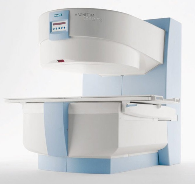
The model under consideration differs from competitors in the form of execution - it is of an open type, and access to the patient is carried out from three sides. The power of the tomograph is 0.2 Tesla, which is one of the smallest values in such MRI. The open form allows a person to sit comfortably, this is also facilitated by a roomy table that can be removed and installed in place. The bed can be rolled back and completely disconnected, it is possible to use 2 tables, thanks to which they conduct studies on patients with increased body weight. The magnet has a C-shape, which is associated with the design of the device.
Users note the ease of use and reliability of the tomograph, as well as the low cost of consumables. Despite the apparent simplicity of design, the model has a large number of complex integrated systems. So, the IPA system makes it possible to change the location of the coils for a full scan of the human body, the InLine system, when collecting the received data, simultaneously corrects the bias, thereby eliminating the misinterpretation of a particular area.
The model is focused on conducting studies of most organs, and is used to study the respiratory, genitourinary system, head, spine, abdominal, chest, etc. It is often used in pediatrics, in some cases even sedation of small patients is not required.
Since the model does not implement cryogenic cooling, its maintenance costs are lower compared to competitors. The manufacturing technology also provides for a long disconnection from the power supply, and therefore the mechanism can be turned off after the end of the work shift. Users also note the possibility of conducting short diagnostic examinations (for example, at one breath hold). The manufacturer claims that on its equipment a slice up to 0.05 mm thick is obtained, in the field of view up to 5 mm. The average price of a product starts from 10,000,000 or more.
- products have a registration certificate and comply with international standards;
- suitable for use in physiotherapy rooms;
- open form;
- low cost compared to high-field products.
- not suitable for complex studies.
Hitachi Aperto 0.4T
votes 12
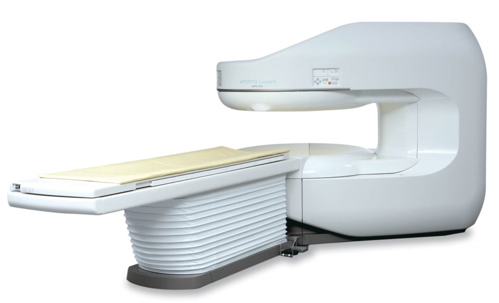
The review continues with the product of one of the best Japanese manufacturers of medical equipment, which is used in many budget clinics. Thanks to the technology used, which takes pictures in an ultra-fast mode, it is possible to conduct diagnostic studies of children without anesthesia, since the possibility of obtaining a "blurred frame" is excluded. Despite the fact that the pictures are taken quickly, they are not inferior in quality to their counterparts.
The field strength is 0.4 Tesla, while the manufacturer claims that the resulting image is not inferior to those made on 1.0 T devices. The device operates in a 320º plane. The table can move in two planes - longitudinal and transverse, making it easy to choose a comfortable position for a person.
The built-in image pre-evaluation system allows you to evaluate their quality and change the settings for taking the next frame. The system of fast image output allows to carry out surgical manipulations, controlling the course of the procedure. Fluoro Triggered CE-MRA technology triggers scanning at the moment when the concentration of contrast agent in the human body reaches its peak value. Most often, studies are carried out in diseases such as diseases of the abdominal organs, spine, joints, head, and chest. The average price of a product starts from 22,000,000 rubles.
- wide functionality - despite the fact that the device is low-field, in many respects it is not inferior to some high-field models;
- suitable for examining children and patients with claustrophobia;
- high resolution.
- there are difficulties with where you can buy a device for free sale - large medical equipment stores rarely work with this manufacturer, and you can order a tomograph from a photo online only in a few online stores.
GE Ovation 0.35 T
votes 7
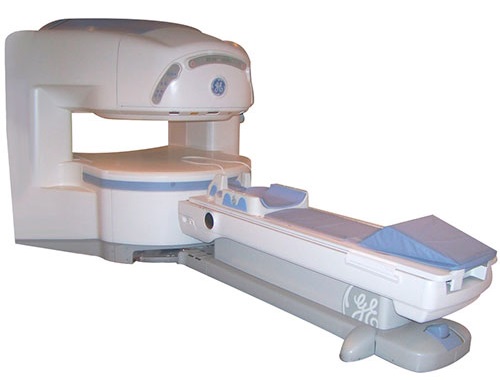
The product of the American company General electric, which also belongs to the category of low-field tomographs, is not inferior to competitors. The field strength is 0.35 Tesla. The scope of the model is the study of the state of the central nervous and cardiovascular systems, spine, ENT organs, joints (including knees). Diagnostics can be carried out on people of any height and weight (from small children to obese), for this, a large number of coil types are included in the package.
The universally shaped, flat and wide magnet is designed in such a way as to leave more free space for the subject, so that the person does not experience discomfort from the closed space. Among the features of the device, several functions can be distinguished. EXCITE - a multi-channel technology is used that collects information simultaneously from several sources and transmits it to the user in an accelerated mode. A large number of coils of different sizes makes it possible to examine not only individual parts of the body, but the human body as a whole. The table for the patient moves in two planes - longitudinal and transverse by 12 cm relative to the center. The average price of a product is 18,000,000 rubles.
- suitable for examining people with claustrophobia and children without sedation;
- fast data processing;
- a large number of settings.
- not the highest quality of the resulting image;
- some buyers complain about how much the tomograph costs - in this price range you can find a model with great functionality.
Voltage 1.5 Tesla
Magnetom Symphony 1.5T Siemens
votes 25
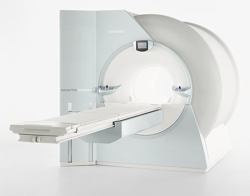
The model of the German manufacturer is well known all over the world due not only to durability and reliability, but also to the price / quality ratio. The voltage in the device is 1.5 Tesla, one magnet is used (weighing 4.05 tons). The scope of the product is cardiology and neurosurgery. Despite the fact that the model first appeared on the market, in terms of its characteristics it is practically not inferior to modern tomographs.
The manufacturer in its products uses the patented technology Maestro Class, which controls the displacement of the position of the human body relative to the initial position, processes the information received during the scanning process, and also speeds up the process of collecting initial data. The intelligent system collects information from 5 streams simultaneously.
According to the clinics that buy such equipment, the device can obtain an image cut from 0.05 mm, even with a small field of view. Users note a simple and intuitive interface. Thanks to individual settings, the doctor can adjust the device to his needs and speed up the routine process of examining patients. Unlike Chinese competitors, the manufacturer of the tomograph in question constantly updates the software and brings it in line with the latest medical technology.
In order to improve data processing speed and throughput, the model can be equipped with an additional console, as well as a workstation, at the request of the buyer. The retractable console is designed in such a way that the patient feels comfortable and does not feel claustrophobic.
The manufacturer also notes the presence of Integrated Panoramic Array technology, which is designed in such a way as to cover the entire body of the subject. There is also a built-in Phoenix system that simplifies the exchange of information between compatible medical devices. To install a tomograph, you will need a room of 30 m2, with a ceiling height of at least 2.4 m. The average price of a used product is 12 million rubles, the cost of a new one is about 40 million rubles.
- there are a large number of used models on the market that can be considered for a small clinic that does not have free cash;
- brand products have the necessary quality certificates;
- the best price / quality ratio;
- since the model is popular, it is easy to find the necessary components and spare parts for it;
- high speed of processing of received images.
- off-budget cost.
Philips Achieva 1.5T
votes 29
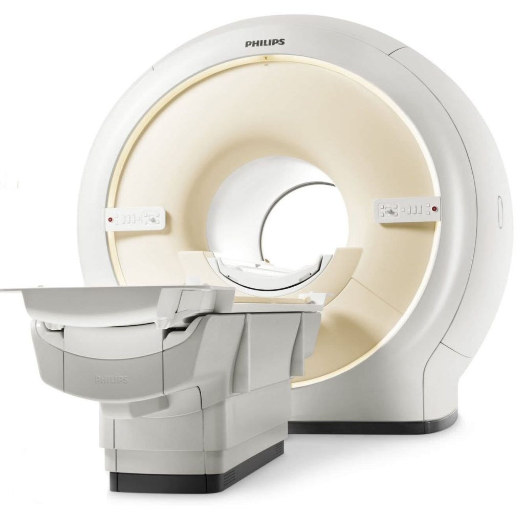
According to radiologists, this is one of the best developments of the Philips brand. Despite the fact that the product has been in production for 10 years, the popularity of models of this type is at its peak. Due to the prevalence of the device, finding spare parts and repair technicians is not difficult. The device is used for the following types of studies: cardiological, spine, head and neck, chest, pelvis, abdominal cavity, lower and upper limbs.
The model is suitable for diagnostic centers that have a room of at least 30 m2 and a ceiling height of 2.65 m. Several advanced technologies are implemented in the product. The main ones are: MultiTransmit (increased scanning speed, individual adjustment for the patient), SmartExam (customizable scanning sequence that starts with one click of the mouse), FreeWave (imaging technology, the feature of which is to display high-resolution images), Sense (parallel imaging multiple images).
The allowable patient weight is 230 kg. These restrictions are related to the diameter of the tunnel (60 centimeters). The company's products are certified, have a registration certificate, and comply with international standards, undergo an annual quality check.Compatible gradient systems - Pulsar, Nova, NovaDual. The number of channels, depending on the tasks set, can be 8, 16, 32. The average price of a new product is more than 30 million rubles, a used device is about 18 million rubles.
- attractive appearance;
- a large number of settings;
- partially closed tunnel allows for research by people with claustrophobia;
- high quality visualization.
- not suitable for brain examination.
Toshiba Vantage Titan 1.5T
votes 24
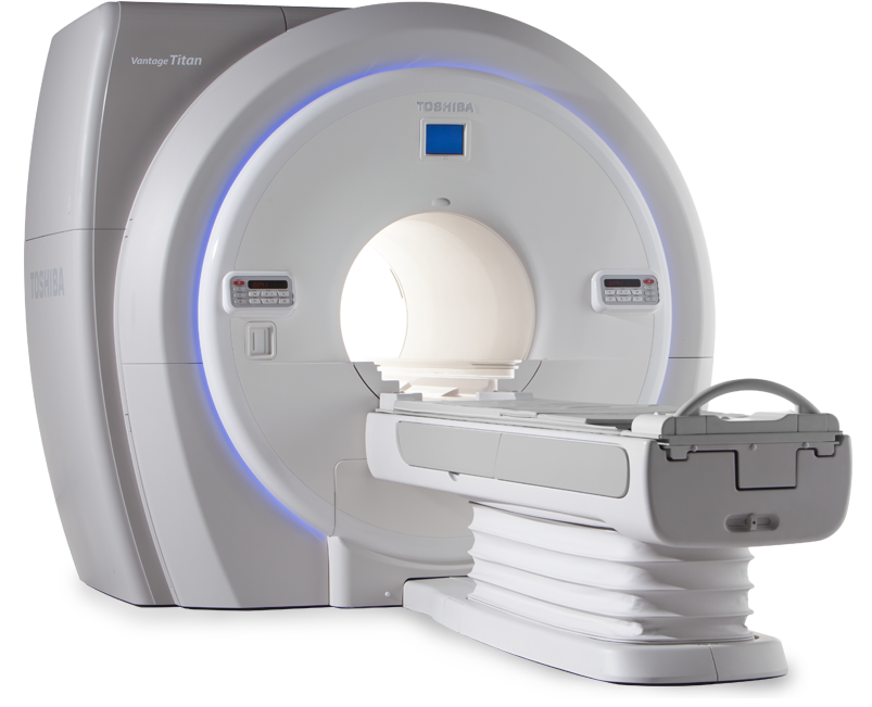
The products of the Japanese company are not inferior to competitors in the quality of their products. Buyers note the low noise level, compact size, short tunnel (149 centimeters), which is combined with a wide internal diameter (71 cm). The semi-open technology allows diagnosing claustrophobic patients as well as children. The product implements Atlas technology, which speeds up research while maintaining the quality of the final image. It is possible to conduct a non-contrast study of blood vessels (arteries and veins).
The manufacturer claims that the tomograph is capable of reproducing images of the entire body of the patient (diffusion-weighted technique). The device examines individual parts of the human body, and then recombines the images into a single whole picture through the use of matrix coils. It is stated that the field of view is the largest among competitors - 50 * 55 * 55 cm. The scope of use is the abdominal cavity, spine, brain and spinal cord, pelvis, vascular system, soft tissues. For diagnostics, 2 channels are used - 16 and 32. The maximum patient weight is 230 kg.
The package also includes a set of spare coils, software, and an instruction manual.The device can be installed in rooms with an area of more than 55 m2. The average price of a new product is 32 million rubles.
- wide and short tunnel, thanks to which patients of any physique can be examined;
- low noise level;
- compact dimensions;
- the possibility of diagnosing the whole body of the patient.
- high price;
- hard to find on the open market.
Ultra high-field
Siemens Magnetom Trio A Tim 3.0T
votes 11
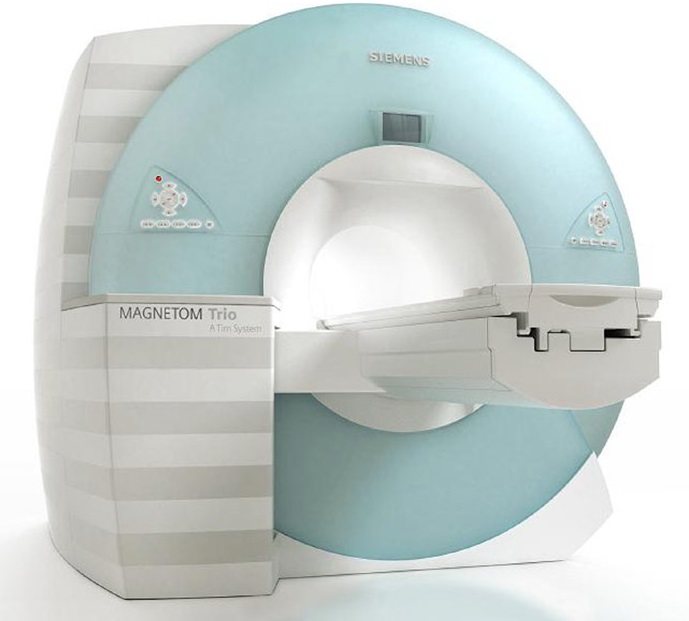
The model under consideration belongs to high-precision mechanisms, and according to the manufacturer's description, it is focused on conducting research work, identifying serious diseases, including oncological ones. The tomograph is in the TOP of the most powerful devices in the world. According to the manufacturer, the mechanism is the most equipped and flexible to perform various tasks.
The user interface with Syngo function provides easy control. The technology of frame processing during scanning speeds up the diagnostic process, since these two processes run in parallel. The Phoenix application independently sets the sequences according to the initial data received from the DICOM image.
The built-in Tim system is designed to have a technological headroom for upgrading, so that after the release of the next update, the device will continue to compete with models from other manufacturers. Buyers note the presence of universal coils that can change their size depending on the needs of the user, so you do not have to buy them separately if necessary.
Since for some patients it is important how the diagnostic equipment looks, when buying the model in question, you can not worry about this parameter - the appearance of the device does not inspire concern. The comfort of the patient during the examination is also provided by the table cover - genuine leather. The average price of a used product is 28 million rubles.
- one of the most technologically advanced tomographs available in clinics;
- the product is included in the TOP of medical equipment, according to the advice and recommendations of rating agencies;
- high accuracy of the received images.
- high price.
Toshiba Vantage Titan 3T
votes 5
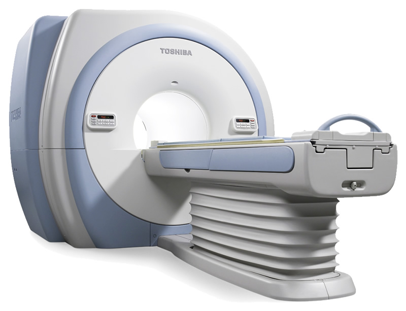
The products of the Japanese brand belong to the category of high-end tomographs, and have a number of advantages. Among them are: a wide diameter of the tunnel - 71 cm, an enlarged field of view - 50 * 50 * 45. Compared to competitors, the noise level in the device is an order of magnitude lower due to the design of the coils, which are placed in vacuum compartments, which makes it possible to reduce the produced sound by more than 90%.
Parallel data processing allows you to simultaneously carry out diagnostics and form a picture, thereby reducing the time spent by a person inside the tunnel. Users note the possibility of conducting an examination without the use of a contrast agent, which takes less time compared to the standard procedure. The entire body can be examined at one time. It is possible to connect up to 128 coils at the same time, some of them are built into the table. It is equipped with a hydraulic drive, moves in all directions, including descending down to a distance of up to 42 cm. The maximum patient weight is 200 kg.
A handy feature for many users is the ability to obtain a large number of sections in a single scan. The equipment is compatible with the international DICOM system. The average price of a used product starts from 32 million rubles, the cost of a new one will cost twice as much.
- wide functionality;
- high image quality;
- a large number of settings;
- low noise level.
- Only a few clinics can afford to buy equipment of this price range.
GE Signa HDxt 3.0T
votes 4
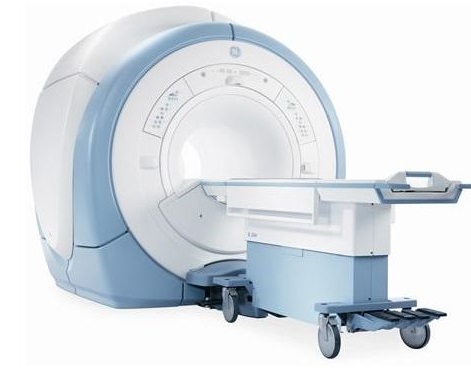
The review is completed by the model of the American representative, which belongs to the “premium” category. The equipment is intended, first of all, for high-precision research, including scientific research, as well as for the detection of various neoplasms, including oncological ones. In addition, it is possible to identify dangerous conditions for human health in the field of cardiology, neurology, and angiology.
The design of the device (8 independent RF channels, refueling with helium once every four years, high-quality components) allows routine maintenance to be carried out less frequently than in equipment from other manufacturers. The magnet also has some features - small size and high uniformity. For the comfort of a person in the tunnel, there is a built-in noise suppression system, which, according to the manufacturer, reduces its level by 40%.
The system is compatible with the DICOM standard, as well as ECG and VCG devices. The table is detachable and moves in any direction, which allows you to put the patient on it in another room, and deliver it to the apparatus. The price of a used product starts from 30 million rubles.
- in the manufacture, the latest technologies were used at the time of development, while the manufacturer supports its products and releases updates;
- detachable table for the patient;
- low noise level;
- universal design.
- difficult to find on sale;
- high price.
Conclusion
When choosing which tomograph of which company is better to buy, it is recommended to focus not only on the financial capabilities of the clinic, but also on the technical characteristics of the product. Such equipment must be purchased only after a mandatory consultation with a technical specialist who will compare the possibilities of the premises and the expected requirements for the equipment with the expected result, and also indicate the estimated cost of maintaining the tomograph, which in some models can cost a significant amount of money.
Some clinics are considering buying only new tomographs that have a high cost. We recommend that you also consider used appliances, as this practice is now widespread. There are a large number of companies that are engaged in the import, restoration and maintenance of such equipment. Due to the fact that most manufacturers of sophisticated equipment support its operation even after a large number of years after manufacture, releasing updates and software for it, some high-end used devices are practically not inferior to new ones, and in some cases even surpass them.
We hope that our article will help you make the right choice!
Popular Articles
-

Top ranking of the best and cheapest scooters up to 50cc in 2025
Views: 131652 -

Rating of the best soundproofing materials for an apartment in 2025
Views: 127691 -

Rating of cheap analogues of expensive medicines for flu and colds for 2025
Views: 124520 -

The best men's sneakers in 2025
Views: 124034 -

The Best Complex Vitamins in 2025
Views: 121940 -

Top ranking of the best smartwatches 2025 - price-quality ratio
Views: 114981 -

The best paint for gray hair - top rating 2025
Views: 113396 -

Ranking of the best wood paints for interior work in 2025
Views: 110319 -

Rating of the best spinning reels in 2025
Views: 105330 -

Ranking of the best sex dolls for men for 2025
Views: 104367 -
Ranking of the best action cameras from China in 2025
Views: 102217 -

The most effective calcium preparations for adults and children in 2025
Views: 102012